New Chemical Tool Developed for Infection Research
08/29/2024
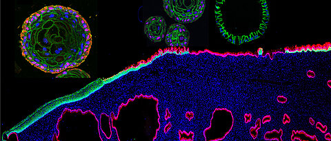
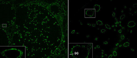
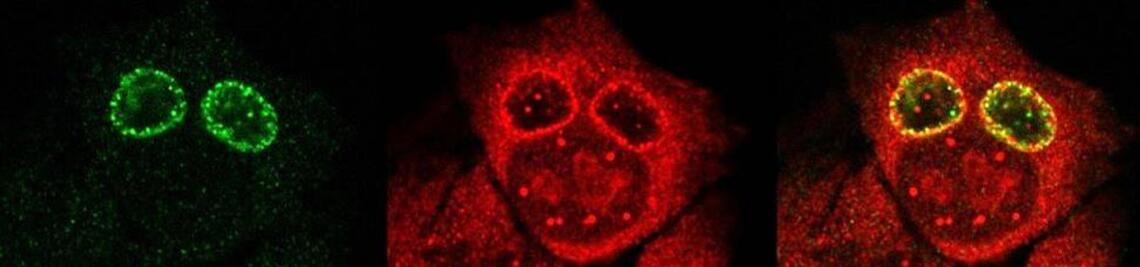
Researchers from Würzburg and Berlin present a new molecule for visualising the sphingomyelin metabolism. This offers prospects for innovative therapeutic approaches in infection research.
more