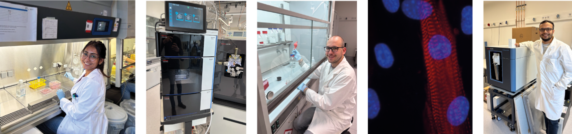
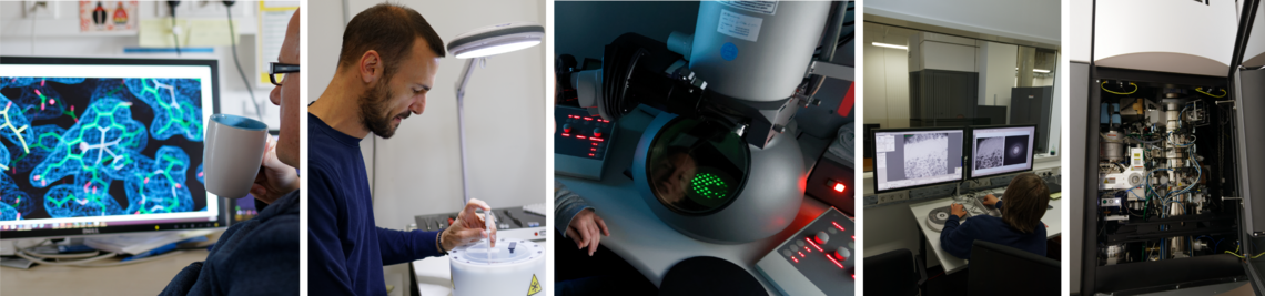
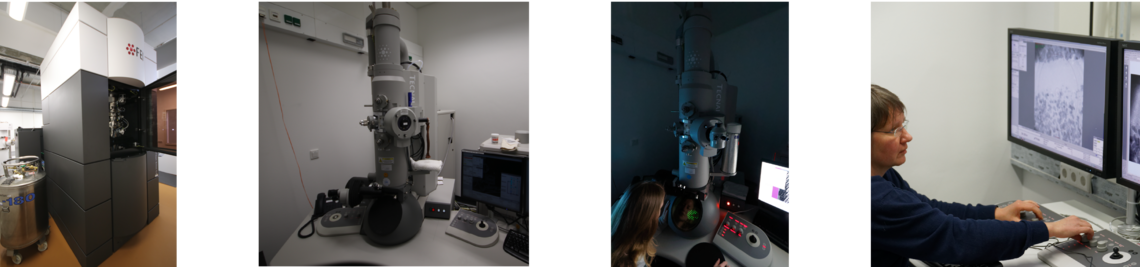
Zentrale Einrichtung Kryo-Elektronenmikroskopie
Erwähnung in Danksagungen
Nehmen Sie die folgende Erklärung in die Danksagung Ihrer Veröffentlichung auf:
„Die Elektronen-Kryo-Mikroskopie wurde in der Kryo-EM-Anlage der Julius-Maximilians-Universität Würzburg durchgeführt, gefördert durch die Deutsche Forschungsgemeinschaft (DFG) - 359471283, 456578072, 525040890.“
Zugang und Kontakt
Der Zugang ist gebührenpflichtig und wird durch eine Zugangsordnung geregelt, die Sie hier herunterladen können.
Wenn Sie an einem Zugang interessiert sind, ein Angebot benötigen oder Ihr Projekt besprechen möchten: Bitte kontaktieren Sie cryo-em@uni-wuerzburg.de
Veröffentlichungen von Nutzern
V. J. Flegler, A. Rasmussen, R. Hedrich, T. Rasmussen, B. Bottcher (2025) Mechanosensitive channel engineering: A study on the mixing and matching of YnaI and MscS sensor paddles and pores. Nat Commun 16: 7881
M. Griessmann, T. Rasmussen, V. J. Flegler, C. Kraft, R. Schneider, M. Hateley, L. Spantzel, M. P. Stevens, M. Börsch, B. Böttcher (2025) Structure of lymphostatin, a large multi-functional virulence factor of pathogenic Escherichia coli. Nature Communications 16
V. Khayenko, C. Makbul, C. Schulte, N. Hemmelmann, S. Kachler, B. Bottcher, H. M. Maric (2025) Induction of hepatitis B core protein aggregation targeting an unconventional binding site. Elife 13
J. Kuper, T. Hove, S. Maidl, H. Neitz, F. Sauer, M. Kempf, T. Schroeder, E. Greiter, C. Hobartner, C. Kisker (2024) XPD stalled on cross-linked DNA provides insight into damage verification. Nat Struct Mol Biol 31: 1580-1588
A. Ullrich, J. Schneider, J. M. Braz, E. Neu, N. Staffen, M. Stanek, J. Blahova, T. Hove, T. Albert, A. Allikalt et al (2025) Discovery of a functionally selective serotonin receptor (5-HT(1A)R) agonist for the treatment of pain. Sci Adv 11: eadv9267
M. Gerovac, K. Chihara, L. Wicke, B. Bottcher, R. Lavigne, J. Vogel (2024) Phage proteins target and co-opt host ribosomes immediately upon infection. Nat Microbiol 9: 787-800
S. Endres, S. Ehrmanntraut, L. Endres, K. Can, C. Kraft, T. Rasmussen, R. Luxenhofer, B. Bottcher, B. Engels and A. C. Poppler (2023) Structural Investigation on How Guest Loading of Poly(2-oxazoline)-Based Micelles Affects the Interaction with Simulated Intestinal Fluids. 10.1021/acsbiomaterials.3c00645
S. Sekulovski, L. Susac, L. S. Stelzl, R. Tampe and S. Trowitzsch (2023) Structural basis of substrate recognition by human tRNA splicing endonuclease TSEN. 30. 834-840.
S. Endres, E. Karaev, S. Hanio, J. Schlauersbach, C. Kraft, T. Rasmussen, R. Luxenhofer, B. Bottcher, L. Meinel and A. C. Poppler (2022) Concentration and composition dependent aggregation of Pluronic- and Poly-(2-oxazolin)-Efavirenz formulations in biorelevant media. J Colloid Interface Sci 606, 1179-1192.
L. T. Alexander, R. Lepore, A. Kryshtafovych, A. Adamopoulos, M. Alahuhta, A. M. Arvin, Y. J. Bomble, B. Bottcher, C. Breyton, V. Chiarini, N. B. Chinnam, W. Chiu, K. Fidelis, R. Grinter, G. D. Gupta, M. D. Hartmann, C. S. Hayes, T. Heidebrecht, A. Ilari, A. Joachimiak, Y. Kim, R. Linares, A. L. Lovering, V. V. Lunin, A. N. Lupas, C. Makbul, K. Michalska, J. Moult, P. K. Mukherjee, W. S. Nutt, S. L. Oliver, A. Perrakis, L. Stols, J. A. Tainer, M. Topf, S. E. Tsutakawa, M. Valdivia-Delgado and T. Schwede (2021) Target highlights in CASP14: Analysis of models by structure providers. Proteins
V. J. Flegler, A. Rasmussen, K. Borbil, L. Boten, H. A. Chen, H. Deinlein, J. Halang, K. Hellmanzik, J. Loffler, V. Schmidt, C. Makbul, C. Kraft, R. Hedrich, T. Rasmussen and B. Bottcher (2021) Mechanosensitive channel gating by delipidation. Proc Natl Acad Sci U S A 118, e2107095118.
A. Grauel, J. Kagi, T. Rasmussen, I. Makarchuk, S. Oppermann, A. F. A. Moumbock, D. Wohlwend, R. Muller, F. Melin, S. Gunther, P. Hellwig, B. Bottcher and T. Friedrich (2021) Structure of Escherichia coli cytochrome bd-II type oxidase with bound aurachin D. Nat Commun 12, 6498.
C. Grimm, J. Bartuli, B. Boettcher, A. A. Szalay and U. Fischer (2021) Structural basis of the complete poxvirus transcription initiation process. Nat Struct Mol Biol 28, 779-788.
F. B. Heiss, J. L. Daiss, P. Becker and C. Engel (2021) Conserved strategies of RNA polymerase I hibernation and activation. Nat Commun 12, 758.
C. Makbul, V. Khayenko, H. M. Maric and B. Bottcher (2021) Conformational Plasticity of Hepatitis B Core Protein Spikes Promotes Peptide Binding Independent of the Secretion Phenotype. Microorganisms 9, 956.
C. Makbul, C. Kraft, M. Grießmann, T. Rasmussen, K. Katzenberger, M. Lappe, P. Pfarr, C. Stoffer, M. Stöhr, A.-M. Wandinger and B. Böttcher (2021) Binding of a Pocket Factor to Hepatitis B Virus Capsids Changes the Rotamer Conformation of Phenylalanine 97. Viruses 13, 2115.
V. J. Flegler, A. Rasmussen, S. Rao, N. Wu, R. Zenobi, M. S. P. Sansom, R. Hedrich, T. Rasmussen and B. Bottcher (2020) The MscS-like channel YnaI has a gating mechanism based on flexible pore helices. Proc Natl Acad Sci U S A 117, 28754-28762.
C. Makbul, M. Nassal and B. Bottcher (2020) Slowly folding surface extension in the prototypic avian hepatitis B virus capsid governs stability. Elife 9, e57277.
K. Stokes, A. Winczura, B. Song, G. Piccoli and D. B. Grabarczyk (2020) Ctf18-RFC and DNA Pol form a stable leading strand polymerase/clamp loader complex required for normal and perturbed DNA replication. Nucleic Acids Res 48, 8128-8145.
J. Wiest, J. Kehrein, M. Saedtler, K. Schilling, E. Cataldi, C. A. Sotriffer, U. Holzgrabe, T. Rasmussen, B. Bottcher, M. Cronin-Golomb, M. Lehmann, N. Jung, M. Windbergs and L. Meinel (2020) Controlling Supramolecular Structures of Drugs by Light. Mol Pharm 17, 4704-4708.
N. Famelis, A. Rivera-Calzada, G. Degliesposti, M. Wingender, N. Mietrach, J. M. Skehel, R. Fernandez-Leiro, B. Böttcher, A. Schlosser, O. Llorca and S. Geibel (2019) Architecture of the mycobacterial type VII secretion system. Nature 576, 321-325.
C. Grimm, H. S. Hillen, K. Bedenk, J. Bartuli, S. Neyer, Q. Zhang, A. Huttenhofer, M. Erlacher, C. Dienemann, A. Schlosser, H. Urlaub, B. Böttcher, A. A. Szalay, P. Cramer and U. Fischer (2019) Structural Basis of Poxvirus Transcription: Vaccinia RNA Polymerase Complexes. Cell 179, 1537-1550 e19.
T. Rasmussen, V. J. Flegler, A. Rasmussen and B. Böttcher (2019) Structure of the Mechanosensitive Channel MscS Embedded in the Membrane Bilayer. J Mol Biol 431, 3081-3090.
B. Song, J. Lenhart, V. J. Flegler, C. Makbul, T. Rasmussen and B. Böttcher (2019) Capabilities of the Falcon III detector for single-particle structure determination. Ultramicroscopy 203, 145-154.
A. Thesseling, T. Rasmussen, S. Burschel, D. Wohlwend, J. Kagi, R. Muller, B. Böttcher and T. Friedrich (2019) Homologous bd oxidases share the same architecture but differ in mechanism. Nat Commun 10, 5138.


