
Impulse for Research on Fungi
04/08/2020For the first time, the cells of fungi can also be analysed using a relatively simple microscopic method. Researchers from Würzburg and Cordoba present the innovation in the journal "Frontiers in Microbiology".
Fungi play an important role for mankind. In the soil, they decompose dead organic material, making it accessible to plants as a nutrient. In industrial biotechnological plants, fungi produce vast quantities of chemicals and food every day. In addition, fungi produce very complex active ingredients that could have potential for medical applications.
On the other hand, there are fungi that damage crops or make people sick. For example, infections caused by the fungus Aspergillus fumigatus can be fatal – especially people with a severely weakened immune system, for example after stem cell or organ transplantation, are affected.
Despite this great importance, far less is known about fungi than about other organisms. "New findings in fungal biology also require that as many researchers as possible have access to state-of-the-art analytical methods," says Dr. Ulrich Terpitz from the Biocenter at Julius-Maximilians-Universität (JMU) Würzburg in Bavaria, Germany.
Successful with expansion microscopy
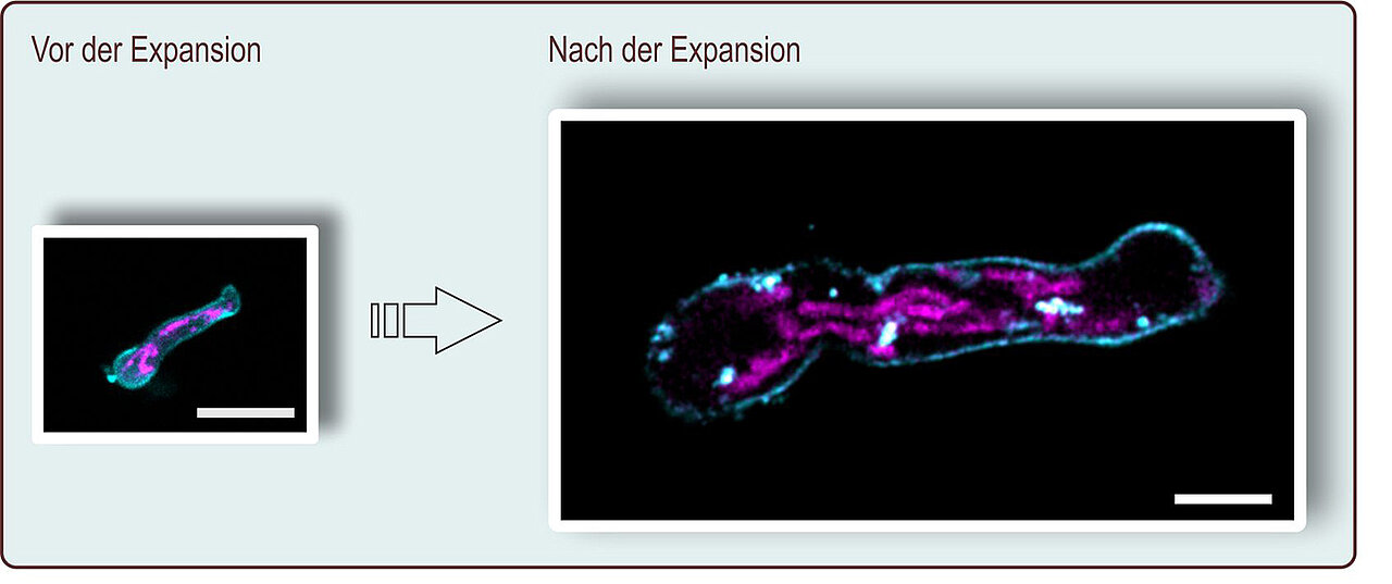
Terpitz's team has developed such a method in collaboration with three other research groups: For the first time, fungi can now be visualised using expansion microscopy (ExM). This method makes cellular structures visible with a resolution of less than 60 nanometres – using a conventional confocal fluorescence microscope.
"Although this is less than can be achieved with high-tech super-resolution fluorescence microscopes, access to such facilities is often limited, especially for fungal researchers. In contrast, standard fluorescence microscopes are widely used and expansion microscopy can be carried out in any biological laboratory," said Terpitz.
The JMU working groups of Professor Markus Sauer (Biocenter) and Dr. Johannes Wagener (Institute of Hygiene and Microbiology) and the group "Molecular Genetics of Fungal Pathogenicity" headed by Professor Antonio Di Pietro from the University of Cordoba in Spain contributed to the development.
Method used for three fungal species
The ExM procedure is quite simple: the stained fungus sample is embedded in a polyacrylamide hydrogel and the fluorescent dyes are anchored in the hydrogel. After water is added, the polymer expands like a gummy bear in a glass of water. It also expands the fluorescent dyes in a uniform manner.
However, there is a problem to be solved first: Fungi have a cell wall that must be removed before expansion. The researchers use cell wall dissolving enzymes for this purpose. "The result is an easy-to-handle protocol for the ExM of fungi that can be used for different fungus species, including the clinically relevant Aspergillus fumigatus," said Terpitz. The researchers also applied their method to the pathogens of corn blight (Ustilago) and tomato wilt (Fusarium oxysporum).
Funding by the German Research Foundation
These results were developed within the framework of the transregional collaborative research centre 124 FungiNet (Jena/Würzburg), which is funded by the German Research Foundation (DFG). Next, the JMU researchers want to use expansion microscopy to show the immune system's reaction to invading fungi. For this purpose, they will bring fungi in cell cultures into contact with immune cells.
Publication
Götz, R., Panzer, S., Trinks, N., Eilts, J., Wagener, J., Turra, D., Di Pietro, A., Sauer, M., and Terpitz, U.; Expansion microscopy for cell biology analysis in fungi. Frontiers in Microbiology, 3rd April 2020, doi: 10,3389/fmicb.2020.00574
Contact person
PD Dr. Ulrich Terpitz, Chair of Biotechnology and Biophysics, University of Würzburg, ulrich.terpitz@uni-wuerzburg.de