Progress in Super-Resolution Microscopy
12/17/2018Does expansion microscopy deliver true-to-life images of cellular structures? That was not sure yet. A new publication in "Nature Methods" shows for the first time that the method actually works reliably.
Immersing deeper and deeper into cells with the microscope. Imaging the nucleus and other structures more and more accurately. Getting the most detailed views of cellular multi-protein complexes. All of these are goals pursued by the microscopy expert Markus Sauer at the Biocenter of Julius-Maximilians-Universität Würzburg (JMU) in Bavaria, Germany. Together with researchers from Geneva and Lausanne in Switzerland, he has now shown that a hitherto doubted method of super-resolution microscopy is reliable.
We are talking about ultrastructural expansion microscopy (U-ExM). In a nutshell, it works like this: The cell structures to be imaged, in this case multi-protein complexes, are anchored in a polymer – just like decorating a Christmas tree.
Cell structures are not distorted
Then the interactions between the proteins are destroyed and the polymer is swelled with liquid. "The polymer then expands uniformly in all spatial directions by a factor of four. The antigens are retained and can subsequently be stained with dye-labeled antibodies," says Professor Sauer. So far, many scientists have been of the opinion that the expansion of the polymer does not proceed uniformly and one gets a distorted representation in the end.
"With U-ExM, we can really depict ultrastructural details, the method is reliable," says Sauer. "And it delivers a picture that is four times higher resolved than with standard methods of microscopy."
Centrioles made the start
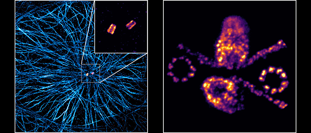
The research team is currently proving this in the journal "Nature Methods" using the example of the centrioles. These cylindrical protein structures play an important role in cell division; the Würzburg biologist Theodor Boveri first described it in 1888.
The centrioles were chosen for the experiment because their structure is already well known. "This enabled us to see, in comparison to electron micrographs, that U-ExM works reliably and even preserves the chirality of the microtubule triplets that make up the centrioles," explains Sauer.
Next, the JMU researchers want to use this method of microscopy to analyze cell structures of which one has not yet had such a precise picture. "These are, for example, substructures of the centrioles, the nuclear pore complexes or synaptonemal complexes. All of them are now accessible for the first time with molecular resolution by light microscopy, "said Sauer.
Publication
“Imaging cellular ultrastructures using expansion microscopy (U-ExM)”, Nature Methods, 17 December 2018, DOI 10.1038/s41592-018-0238-1
Contact person
Prof. Dr. Markus Sauer, Chair of Biotechnology and Biophysics,
Biocenter, JMU, T +49 931 31-88687, m.sauer@uni-wuerzburg.de