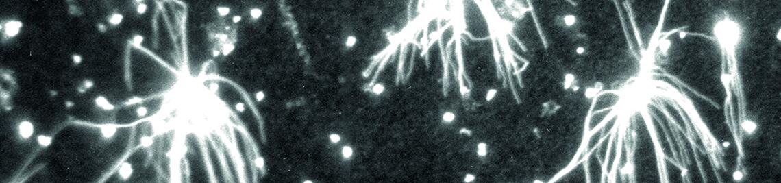
Aster formation in vitro
This image shows isolated centrosomes (bright dots) as nucleators of microtubule „rays“ (fibres) in a cell-free system (immunofluorescence microscopy with anti-tubulin antibodies; photo: Marie-Christine Dabauvalle).
This image shows isolated centrosomes (bright dots) as nucleators of microtubule „rays“ (fibres) in a cell-free system (immunofluorescence microscopy with anti-tubulin antibodies; photo: Marie-Christine Dabauvalle).