Showcase to "Marcella Boveri. Pioneer for women in academia and science"
Theodor Boveri's private microscope with engraved name „TH. BOVERI“ on the blackened base (Ernst Leitz Wetzlar, stand Ia, serial number 10966, 1887)
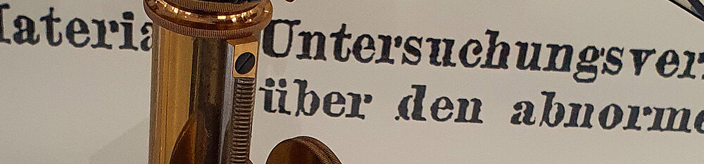
Theodor Boveri acquires this microscope from the Ernst Leitz company in Wetzlar in 1887, when he works at the Zoological Institute of the University of Munich. The attached camera lucida (Zeichenapparat) was developed by Ernst Abbe (1840–1905) and manufactured by various companies. The model shown was made by Bausch & Lomb Optical Co., Rochester N.Y., USA. The drawing apparatus consists of the large, adjustable mirror and a prism system that is fixed above the eyepiece of the microscope and enables the simultaneous view of object and drawing. By placing a drawing paper next to the microscope and looking through the eyepiece with the prism, the image of the specimen appears superimposed on the drawing paper. In this way, the specimen can be traced to scale without taking the eyes off the microscope.
Storage box for 100 microscope slides by Theodor Boveri (replica by Ulrich Scheer)
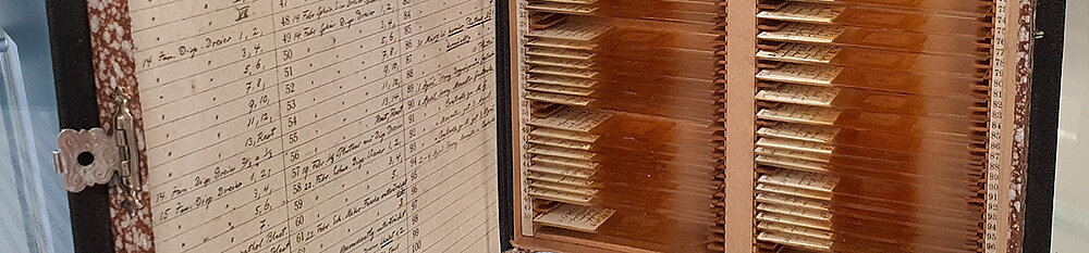
This book-like storage box is entitled of Theodor Boveri's handwritten note „Naples 1901/02, experiments on echinoid eggs“ on the back. He and his wife Marcella prepare the slides in the course of their famous dispermy experiments with sea urchin eggs, which they carry out during their winter stay at the Naples Zoological Station in 1901/02. The slides hold all the information on which Boveri built his chromosome theory of heredity. They are labelled and dated by Boveri. On the inside of the box lid is a handwritten list of all the slides contained. Four such boxes exist with a total of 455 microscope slides.
Closed folder for the storage of 20 microscopic slides used by Theodor Boveri
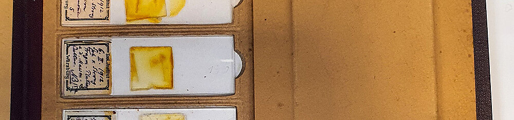
Microscope slides with inscriptions by Marcella Boveri (replica by Ulrich Scheer)
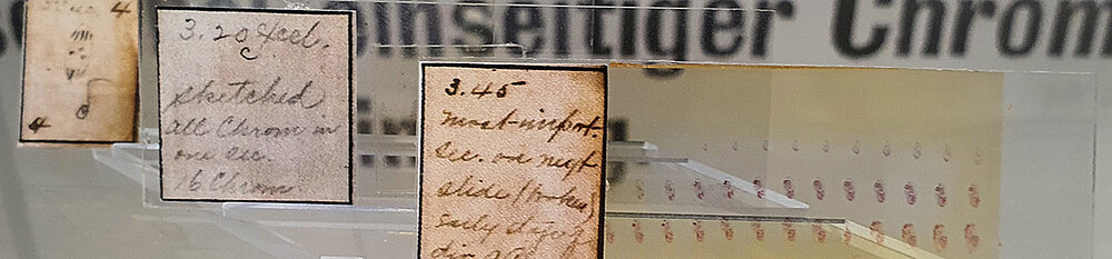
These three slides contain sections of sea urchin embryos. Marcella Boveri handwrites probably in 1902 on it:
• „3.45 most-important. See on next slide (broken) early stage of div[ision] of chrom[osomes].”
• „3.20 […] sketched, all chrom[osomes] in one sec[tion]. 16 Chrom[osomes].”
• „2nd row 5th section”, below sketch of a mitosis.