dStorm / SIM / LSM / Lattice Lightsheet / RCM
Lattice Lightsheet
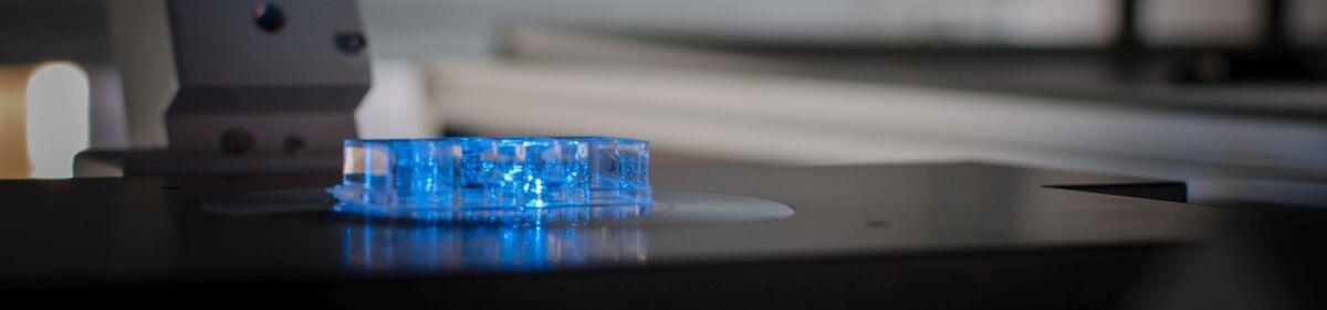
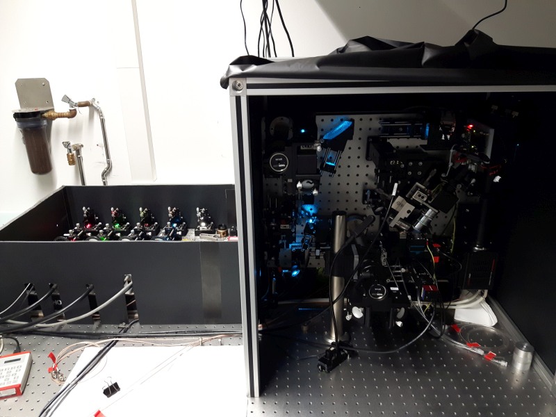
We installed a Lattice Light-Sheet Microscope designed by Eric Betzig, Janelia Farm Research Campus. Light-Sheet Microscopy enables increased imaging speed while keeping photobleaching and phototoxicity to a minimum by illuminating only the focal plane of the detection objective. In Lattice Light-Sheet Microscopy, a lattice of Bessel-Gauss beams is used to create a thin, yet long light-sheet to confine the illumination in the axial direction of the detection objective. This results in unprecedented spatiotemporal resolution when imaging live samples from cells to tissues in 3D over time.
Reference: Chen et al., 2014
| Detection Objective: | Nikon 25XC Apo W Water Dipping |
|---|---|
| Camera: | Hamamatsu sCMOS Orca Flash V3 |
| Available Lasers: | 404 nm, 488 nm, 532 nm, 561 nm, 647 nm |
Two-color live-imaging of COS-7 cells unsing Lattice Light-Sheet microscopy. Microtubules (red) are labeld via SiR-tubulin (Spirochrome) and nucleic acids (blue) are stained with SYTO® 16 (Thermo Fisher Scientific). Four volumes per minute were recorded.
Lattice Light-Sheet microscopy of Fibroblasts labeled with pHrodo™ Red (green) and Shiga-Toxin-Alexa 647 (red) to visualize intracellular trafficking pathways. Two volumes per minute were captured in medium without phenol red.


