Collaborative Research Center / Transregio 166
High-end light microscopy elucidates membrane receptor function
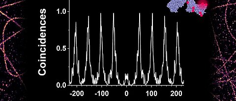
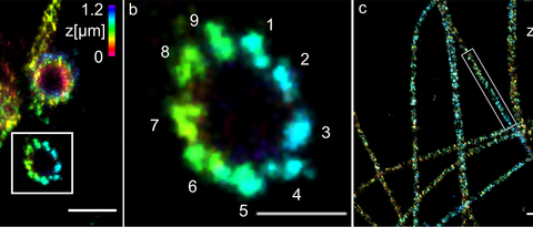
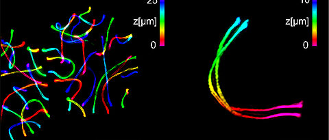
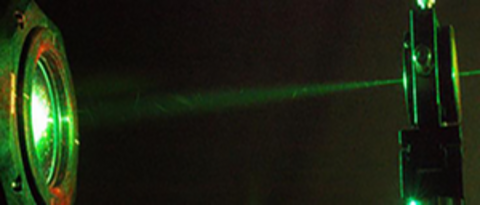
In the CRC/TRR 166 ReceptorLight high-end light microscopy techniques with highest spatial and time resolution are applied and further developed to gain deeper insight into the function of membrane receptors. The participating groups in Jena and Würzburg bundle their methodological expertise in the field of super-resolution microscopy, electrophysiology, and biophysics of membrane receptors to generate new insights into the function and distribution of diverse membrane receptors, and in parallel, to induce the development of new high-end light-microscopy methods.
For more informations see receptorlight
Center for Personalized Molecular Immunotherapy |
|

The European Structural Fund supports two new projects of the University of Würzburg with more than EUR 4.3 million. In close cooperation with the University Hospital and regional companies, research activities aim to drive medical progress.
"The Bavarian State Ministry for Education, Science and the Arts funds two seminal projects which the University of Würzburg runs jointly with regional companies by providing more than EUR 4.3 million in total from funds of the European Regional Development Fund", Bavarian Minister of State, Dr Ludwig Spaenle, announced a few days ago. more >>
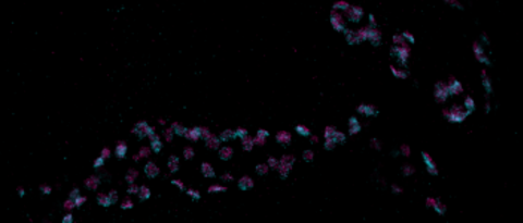
Light-induced cell damage in live-cell super-resolution microscopy
Super-resolution microscopy can unravel previously hidden details of cellular structures but requires high irradiation intensities to use the limited photon budget efficiently. Such high photon densities are likely to induce cellular damage in live-cell experiments. more >>
Latest Publications
-
Decoding the molecular interplay of CD20 and therapeutic antibodies with fast volumetric nanoscopy. . In Science, 387(6730), S. eadq4510-. American Association for the Advancement of Science, 2025.
-
Small fibre neuropathy in Fabry disease: a human-derived neuronal in vitro disease model and pilot data. . In Brain Communications, 6(2), S. fcae095-. 2024.
-
LGI1 Autoantibodies Enhance Synaptic Transmission by Presynaptic Kv1 Loss and Increased Action Potential Broadening. . In Neurology Neuroimmunology & Neuroinflammation, 11(5), S. e200284-. Wolters Kluwer, 2024.