Deutsch
THE BIOCENTER OF THE UNIVERSITY OF WÜRZBURG

Archive

The distribution of the glutamate receptor mGluR4 and other proteins in the presynaptic membrane Left a high-resolution dSTORM image. On the right, the result obtained with conventional fluorescence microscopy – molecular details are not visible here.

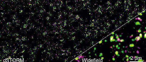
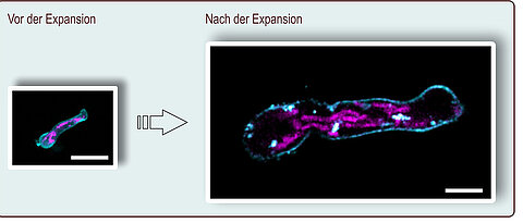
"Distance keeping" is not exactly the motto of the glutamate receptors: Using high-resolution microscopy, it now was discovered that the receptors usually appear in small groups at the synapses and are in contact with other proteins.

more
In expansion microscopy, the preparation is magnified more than four times. Here, a germ tube of Aspergillus fumigatus is shown before and after expansion; the scale corresponds to ten micrometers. The plasma membrane (turquoise) and the mitochondria (pink) were stained. (Picture: Ulrich Terpitz / University of Würzburg)

For the first time, the cells of fungi can also be analysed using a relatively simple microscopic method. Researchers from Würzburg and Cordoba present the innovation in the journal "Frontiers in Microbiology".

more