
dStorm / SIM / LSM / Lattice Lightsheet / RCM
Re-scan Confocal Microscopy (RCM)
We set up a correlative system using the super-resolution technique dSTORM and Re-scan Confocal Microscopy (RCM), which is a resolution-enhanced form of LSM. By re-scanning the emission light after the pinhole, RCM gains a resolution improvement of 1.4 fold. This will increase the lateral resolution from 240 nm of a confocal microscope to 170 nm of the RCM, while maintaining the sectioning capability of a confocal system. Since the detector (Photomultiplier Tube) is replaced by a sCMOS camera with high quantum efficiency, the signal-to-noise ratio of the RCM is twice as good as for a confocal microscope. With this system we are now capable of quantitatively investigating protein distributions in the context of dynamic processes with high spatial resolution.
Reference: de Luca et al., 2013
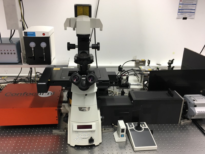
| Microscope-body: | Nikon TiE with motorized stage and PFS-System (Perfect-Focus-System), inverted |
|---|---|
| Objective: | 100x Oil, NA 1.49 Apo-TIRF 60x Water, NA 1.27 100x Siliconoil, 1.3 |
| dSTORM Detection | |
| Laser: | 405nm, 488nm, 640nm: 170mW, 532nm: 500mW |
| Camera: | EMCCD, Andor iXON DU-897 |
| RCM | |
| Laser: | Cobolt Skyra, Multiline lasersystem 405nm, 488nm, 561nm, 640nm: 50mW |
| Camera: | sCMOS, Andor Zyla 4.2P |
 tagged with YFP")
, actin (Phalloidin 488) and microtubuli (Alexa Fluor 647)")


