Groups
RESEARCH GROUPS
Alsheimer
We currently study the function of nucleocytoplasmic network systems in nuclear shaping and meiotic chromosome dynamics
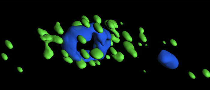
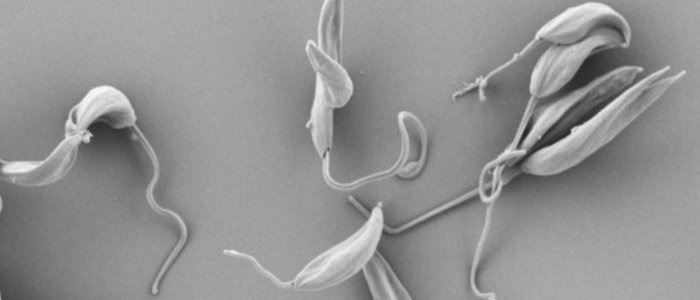
Engstler
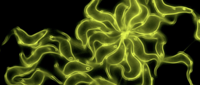
We study the adaptive and dynamic pleomorphism of parasites on the molecular, cellular and organismic level
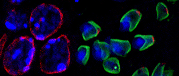
Janzen
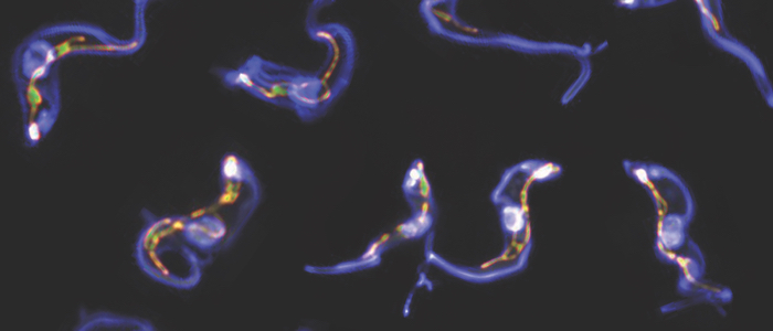
We investigate how African trypanosomes and Leishmania parasites adapt to different host environments