Staff scientists
Manfred Alsheimer
manfred.alsheimer(at)uni-wuerzburg.de
Tel +49 (0)931 31-84282
Room C104
Students anytime (just e-mail me)
Tom Beneke
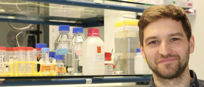
tom.beneke@uni-wuerzburg.de
Websites: LeishGEdit & LeishBASEedit
Room C145
Students anytime (just e-mail me)
Markus Engstler
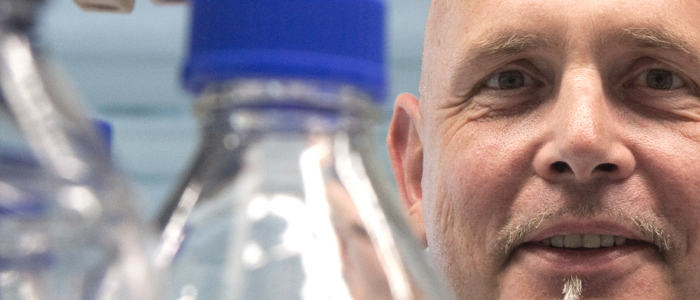
markus.engstler(at)uni-wuerzburg.de
Tel ++49 93131 84250 (PA)
Tel ++49 93131 80060
Skype mengstler
Room C143
Students anytime (just e-mail me)
Christian Janzen
christian.janzen(at)uni-wuerzburg.de
Tel ++49 93131 86685
Room C121
Students anytime (just e-mail me)
Nicola Jones
nicola.jones(at)uni-wuerzburg.de
Tel +49 (0)931 31-88713
Room C127
Students anytime (just e-mail me)
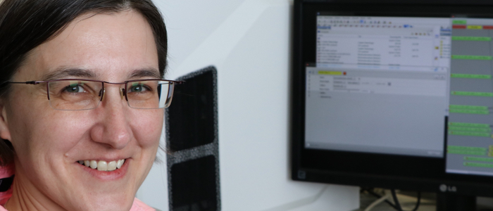
Susanne Kramer
susanne.kramer(at)uni-wuerzburg.de
Tel ++49 93131 86785
Room C147
Students anytime (just email me)
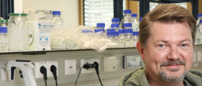
Timothy Krüger
tkrueger(at)biozentrum.uni-wuerzburg.de
Tel ++49 93131 84277
Skype
Room C133
Students Tue, 4-6 p.m.
Christian Stigloher

christian.stigloher@uni-wuerzburg.de
Tel ++49 93131 89850
Room DK16


