Lehrstuhl für Zell- und Entwicklungsbiologie

benavente lab

We investigate the molecular organization, function and evolutionary history of the mammalian meiotic chromosome

Research synopsis

Meiosis is a special type of cell division that is restricted to the germ line. It consists of two successive cellular divisions that take place without intervening DNA replication. Meiosis is the basis for sexual reproduction in that it enables the generation of haploid cells. Furthermore, it represents the largest source of genetic variability. Errors in meiosis are a major cause of aneuploidy and fertility problems also in humans.

During the extended meiotic prophase of the first meiotic division major changes in nuclear architecture take place that are critical for the meiotic process. At early prophase (leptotene stage), homologous chromosomes attach their telomeres to the nuclear envelope. Subsequently, homologous chromosomes, which initially occupy different nuclear territories, move and initiate stable pairing between them (zygotene stage) via a structure called synaptonemal complex (SC). When completely assembled at pachytene stage, each SC extends all along a chromosome bivalent. During diplotene and diakinesis, the SC disassembles and the homologous chromosomes desynapse.

SCs are evolutionarily conserved meiosis-specific karyoskeletal structures which play a key role during synapsis of homologous chromosomes, chiasmata formation and chromosome segregation. SCs show a tripartite ladder-like organization characterized by the presence of two lateral elements (LEs) to which chromatin loops of the individual homologous chromosomes are attached. The third component - the central region (CR) - holds the SC together and is located in the gap between LEs. The CR is composed of a central element (CE) and numerous perpendicularly-oriented transverse filaments (TFs) that link the LEs with the CE. The major protein components of mammalian SCs have been already characterized: the meiosis-specific proteins SYCP1, 2 and 3. More recently, CE-specific proteins have been identified (i.e. SYCE1, 2, 3, 4 and 5).

In order to investigate the molecular architecture and dynamics of the synaptonemal complex, we are applying super-resolution imaging and electron microscopic tomography to meiotic chromosomes and their telomeres of wildtype and selected knockout mice.

Selected publications

Zwettler, F., M.C. Spindler, S. Reinhard, T. Klein, A. Kurz, R.  Benavente* & M. Sauer*: Tracking down the molecular architecture of  synaptonemal complexes with expansion microscopy. Nat. Commun. 11:3222(2020).

Spindler, M.C., J. Redolfi, F. Helmprobst, P. Kollmannsberger, C. Stigloher & R. Benavente: Electron tomography of mouse LINC complexes at meiotic telomere attachment sites with and without microtubules. Commun. Biol.2: 376 (2019).

Dunce, J.M., A.E. Milburn, M. Gurusaran, I. da Cruz, L.T. Sen, R. Benavente* & O.R. Davies*: Structural basis of meiotic telomere attachment to the nuclear envelope by MAJIN-TERB2-TERB1. Nat. Commun. 9: 5355 (2018).

Schücker, K., T. Holm, C. Franke, M. Sauer & R. Benavente: Elucidation of synaptonemal complex structure by super-resolution imaging with isotropic resolution. Proc. Natl. Acad. Sci. USA 112: 2029-2033 (2015).


More...

https://scholar.google.com/citations?view_op=list_works&hl=en&user=gLJI5DgAAAAJ

RESEARCH TOPICS

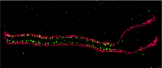
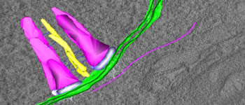
Molecular Architecture of the Meiotic Chromosome

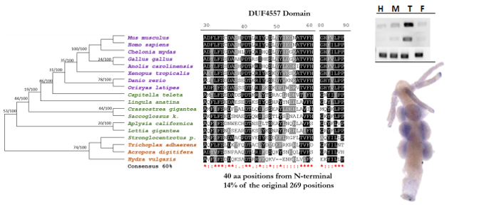
Evolutionary History of Meiosis


Meiotic Gene Expression

A collaborative project with colleagues of the Clemente Estable Institute for Biological Research (Uruguay) to elucidate stage-specific gene expression of meiotic cells

Architecture and Function of the Nuclear Envelope

Roberta Sciurano

Visiting scientist

Fernanda Trovero

Visiting scientist